Lab#1 IntroductiontotheMicroscopy, ObservationofProkaryoticandEukaryotic Cellular material Introduction Manyofthecellsandorganismsthatyouwillbestudyingareatthelowerlimitsofvisibilityoflightmicroscopes, therefore , itisextremelyimportantthatyouattaincriticallightinganddirecting. Itisalsoimportanttohandlethemicroscopecompetentlytoavoiddamagingeitherthemicroscopeorthepreparationyouarestudying. Evenstudentswhohavepreviouslyusedmicroscopesshouldreadtheinstructionscarefully.
GuideBiolabo By using a web rowser, go tothefollowingwebweb page: http://salinella. bio. uottawa. ca/biolabo/(youcantryitfromhome). UnderMicroscopyyouwillfindlinkstopagesthatdescribebothtypeofmicroscopesyouwillusethissemester, aswellashowtosetupandusethese people. Itisstronglyrecommendedthatyouvisitthesepagespriortoattendingyourfirstlab. ImageJ/Qcapture Althoughyoucanmakeallyourobservationsbywatchingdirectlythroughtheoculars, italsocanbedoneonthecomputerscreenusingthedigitalcameraattachedtoeachmicroscopic lense.
Forthat, youwillusetheImageJprogramtogetherwithacaptureplugin calledQcapture. VisitthelabwebsitetolearnhowtouseImageJ(linkonthehomepage). Allobservationscanbemadeonyourcomputerscreenorintheoculars. Eachmethodhasitsadvantagesanddrawbacks, youwillhavetochoosewhichoneitmoreappropriate(ortheoneyouprefer): Oculars Screen? Greaterresolution? Widerfieldofview? Canshareobservationwithothers? Morecomfortableforusers? Takepictureswhileobserving Lab1 Microscopy TheCompoundMicroscope OntheGuideBiolabopageclickontheCX41CompoundMicroscopelinkthenonPartsandFunction. Thiswillbringupalabelledlinediagramofyourmicroscope. Familiarizeyourselfwiththevariouscomponentsshowninthisfigure. After that, clickonSetupandBrightfieldalignmentinordertoknowhowtouseandhandlethemicroscope. Now, locateyourcompoundmicroscopeinthecupboardbelowthesinkofyourworkstation. Placeitonthecounterbetweenthe omputerandtheendofthecounter. Besurethatwheneveryoutransportthemicroscope, itisalwayskepterect, theocularlenswillfalloutifthescopeistiltedorthrown. Eventhoughyoudon’tneedthedissectingmicroscoperighttoday, takeitoutofthecupboardandinstallitbesidethecompoundmicroscope. Connectonefirewirecabletoeachofthecamerasinstalledontopofthemicroscopes. Thisway, everythingissetupforfurtherobservationsbothonyourcomputerscreenandthroughtheoculars. Partsofthecompoundmicroscope
Themicroscopeconsistsofasystemoflenses, alightsource, andagearedmechanismforadjustingthedistancebetweenthelenssystemandobjectbeingseen. Thereareanumberofimportantcomponentsanditisessentialthatyoubeabletoidentifythemandunderstandtheirfunctionbeforeyoucanproceed. BygoingthroughthedifferentmodulesinBiolaboandusingthemicroscopeyouwilldevelopacompetencyforbrightfieldmicroscopy. IdentifythefollowingcomponentsusingBiolabo(Partsandfunctionsfigure)andyourmicroscope:
REVOLVINGNOSEPIECE: Supportsthevariousobjectives Youwillonlyusethe4x, 10xand40xobjectivesintheBIO1140labs(notthe100x). STAGE: Supportsthespecimenbeingobserved. AsystemofknobsonthesideofthestageallowsyoutomovethespecimenundertheobjectiveontheXandYaxes. Tryandmovethestage. COARSEFOCUSCONTROL: Permitsrapidchangeindistancebetweenthespecimenandtheobjectivetherebyallowingforroughfocussing”Donotusewhenfocusingwiththe40xobjective
FINEFOCUSBUTTON: Permitssmallchangesindistancebetweenthespecimenandtheobjectiveandtherebyallowsforfinalfocussingoftheimage. 15 Lab1 Microscopy OCULAROREYEPIECE: Amagnifyingelementinthemicroscopic lense, usually10X. Itisthroughtheocular, oreyepiecethatonelooksatthespecimen. Allourmicroscopesareparfocal, sothatwhenanobjectisinfocuswithoneobjective, thefocuswillnotbecompletelylostwhenchangingtothenextobjective. AIMS: Themagnifyingelementwhichisclosesttothespecimen.
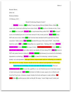
Seefigure1tofindoutabouttheengravingsonthesideofeachobjective. CONDENSER: Systemoflensesthatconcentratesthelightfurnishedbytheilluminator. Itdoesnotmagnifytheobject. CONDENSERHEIGHTADJUSTMENTKNOB: Allowsonetofocustheconcentratedlightontothespecimen. APERTUREIRISDIAPHRAGM: Usedtoreduceglarefromunwantedlightbyadjustingtheangleoftheconeoflightthatcomesfromthecondenser, ProductionofImagebyaCompoundMicroscope Themostimportantpartofamicroscopeisthetarget.
Alltheotherpartsoftheinstrumentaredesignedtohelptheobjectiveproducethebestpossibleimage. Thebestimageisnotthelargest, itistheclearest. Thereisnovaluetoahighmagnifying. Iftheresolutionispooryouwillhavenobetterunderstandingofthespecimen. lightbeam ocularlens Magnification Numericalaperture(NA) Determinestheresolving poweroftheobjective* Opticaltubelength/max. coverslipthicknessinmm prism objectivelens specimen condenserlens Figure1: Objectivesengravings lightsource
Figure2: Imageproductioninacompoundmicroscope. 14 Lab1 Microscopy *Resolvingpoweristheabilitytoseetwoobjectsthatareverycloseastwoseparateobjects. Thehumaneyewillresolvingpowerisabout100m. Usingthecompoundmicroscope AlwayshandlethemicroscopeGENTLY! Itisanexpensive, delicateandheavytool. Carryitwithtwohands, onehandonthearm, andtheotherhandunderthebase. Iftheocularorobjectiveisdirty, wipeitcleanusingONLYKimwipesorspeciallenstissueandcleaningfluidsupplied.
Ifyouuseanythingelseyoumayscratchthelens. Wipeupanycleaningfluidimmediately, otherwiseitwilldissolvethegluewhichholdsthelensinplace. REMEMBER, yourdemonstratorisheretohelp, so, INQUIRE! 1 . Makesurethatthepowercordispluggedintothebackofyourmicroscopeandintoapoweroutlet. 2 . Usingtheletter”emicroscopeslideprovided, followsteps2through13intheSetupandBrightfieldalignmentprocedureofBiolabo. Remember, observationcanbedoneonscreenorthroughtheoculars. Orientationandworkingdistance. Startingyourexaminationwiththe4Xobjective, positiontheletter”e”slideonthestage. 2 . Drawwhatyouseeinthemicroscope: _________________ 3. Whatwouldaslidewiththeletter”tlooklikeunderthemicroscope? _________________ 4. Usingtheknobslocatedonthesideofthestageandlookingthroughthemicroscopic lense, movetheslideslowlytotheright, thentotheleft. Recordyourobservations. ___________________________________5. Today, movetheslideslowlyawayfromyou, thentowardsyouwhileobservingthroughthemicroscope.
Recordyourobservations____________________________________ 6. Focusontheslideat10X. Checkthedistancebetweentheobjectivelensandyourslide(=theworkingdistance, seealsothereferenceattheendofthischapter). Nowswitchtothe40Xobjectiveandchecktheworkingdistance. Whathappenstotheworkingdistanceasyourmagnificationimproves? 12 Lab1 Microscopy Depthoffield(depthoffocus) Lenseshaveadepthoffocus. Itisthenumberofplanesinwhichanobjectappearstobeinfocus.
Extendyourfistatarm’slengthinfrontofyouandholdyourthumbup. Concentrateonyourthumbandnoticethattheobjectspastyourthumbontheothersideoftheroomarenotclearlyseen. Similarlywithamicroscope, whenitisfocussedononesurface, thesurfaceslowerorhigherwillbeoutoffocus. 1 . Positionapreparedslidewithcolouredthreadsuponthestage. Atlowelectrical power, 4X, focusontheareawherethethreadscross. 2 . Usingthefinefocusadjustment, focusupanddownslowly. 3. Repeatusingdifferentaims.
Whatcanyousayaboutthedepthoffieldatdifferentmagnifications? Hasitincreasedordecreased? (i. e., canyouseemorethreadsinonefocalplaneat4Xor40X? ) ____________________________________________________________ Magnification Themagnificationgivenbyobjectivesandocularsisengravedonthem. Thetotalmagnificationforanycombinationofobjectiveandocularistheproductofthemagnificationofeachlens. Objectivemagnification Ocularmagnification TotalMagnification Lightintensity Workingdistance 4x 10x 40x Large 22mm 10x 10x 100x
Medium 15. 5mm 40x 10x 400x Low zero. 56mm Table1 Comparisonzoom, workingdistanceandbrightnessatthreedifferentobjectivemagnifications. Youalsocancalculatethemagnificationofyourpictureusingthefollowingformula: Magnificationfactor=measuredsizeofobject=(X) Actualsizeofobject 13 Lab1 Microscopy SpecimensizeandMagnificationof thepicture Beforeyoustartthisexercise, makesureyouhavecarefullyreadthewebsitesectionrelevanttothesoftwareyouwillusetotakedigitalpictures(ImageJ/Qcapture).
Thegoalofthissectionistoteachyoudifferenttechniquesthatwillallowyoutodeterminethesizeofobjectsyou’reobservingunderthemicroscopic lense. Thegeneralprincipleisfairlybasic: 2objectshavethesamerelativesize(expressedasaratio)intherealworldandunderthemicroscopic lense. actualsizeofobjectA=on? screensizeofobjectA A1=A2actualsizeofobjectBon? screensizeofobjectBB1B2 Thefollowingexercisesareapplicationsofthisformula. Placeaslideunderthemicroscope.
Choosetherightobjectiveandadjustthefocusandlightlevel. In that case, chooseastructureyouwanttomeasureandtakeapicture. A? Firsttechnique: Measuringanobjectusingthefieldofview(FOV): Thesimplestwaytodeterminethesizeofanobjectistousetheknownsizeofthewholefieldofview(FOV, thewholepicturefromlefttoright). 1? Onthecomputerscreen(usingarulerandwithoutwritinganythingofthescreen), measuretheobjectofwhichyouwanttodeterminethesize(=A2) 2? Then, measurethewidthofthewholepictureonthescreen(=B2).? Refertotable2onpage20toknowtheactualsizeofthefieldofviewfortheobjectiveyou’reusing(=B1) 5? Usethefollowingformula: Actualsizeoftheobject(A1)=ActualsizeoftheFOV(B1)xon? screensizeoftheobject(A2) in? screensizeoftheFOV(B2) Case: Onasnapshotusingthe4xobjective, aninsecthasanabout? screenlengthof10cm. Thewholepictureis20cmwide. Whatistheactualsizeoftheinsect? ______________________________ 18 Lab1 Microscopy B? Secondmethod: Measuringanobjectusingascalebarfile:
FromImageJ(usingthefile/opencommand), openthefilethatcontainstherelevantscalebarinthe(T: /BIO/BIO1140): new10X. jpgforthe10xobjective, andnew40X. jpg(forthe4xand40xobjectives). In that case, usingarulermeasurethefollowingdistancesdirectlyonthecomputerscreen: 1? Theon? screenlength(orwidth)oftheobjectwhosesizeyouwishtodetermine(=A2) 2? Thewidthofthescalebaronthescreen(=B2)Younowcancalculatetheactualsizeoftheobjectusingtheformula: actualsizeofobject=on? creenlengthofobjectxactualsizeofscalebar*on? screenlengthofscalebar? A1=A2xB1 B2 *Theactualsizeofthescalebarisindicatedonthescalebarfile(ex: onthenew10x. jpgfile, thebarrepresents0. 2mmat10xor0. 02mmat100x)=B1 Example: Itookapictureofasmallinsectlarva, usingthe4xobjective. Thelarvalengthis60mmonthescreen. Thescalebaronthenew40x. jpgis30mmandrepresents0. 2mm. Whatistheactualsizeofthelarva? _________________________
Donotputthecompoundmicroscopebackinthecupboardyouwillneeditlaterthisafternoon. Pointstorememberconcerningmicroscopes 1 . Alwaysworkwithacleanmicroscope. Useonlythelenspaperprovided. Don’tforgettocleantheslidetoo! 2 . Alwayslocatethespecimenunderlowpowerandworkyourwayuptothehighpowerobjective. several. Neverusethecoarsefocusingknobwhenthehighpowerlensisinlocation. Useonlythefinefocusknob. four. Neverusethe100xin1styearlabs(wedidn’tteachyouhow)5.
Alwaysreadjustilluminationwheneveryouchangethegoal. Toomuchlightwillgiveyouablurryimagethatyoucannotfocuson. 15 Lab1 Microscopy Thestereoscopicmicroscope (dissectingmicroscope) Thestereoscopicmicroscope, alsocalledstereoscopeordissectingmicroscope, isusedtoviewobjectsthataretoolargeortoothicktoobserveunderthecompoundmicroscope. Stereomicroscopesarealwaysequippedwithtwoocularsproducingastereoscopicorthree? dimensionalimage. Unlikethecompoundmicroscope, theimageisnotinverted.
Ourstereomicroscopesprovidemagnificationintherangeof6. 7X 45Xusingazoom? typelensprogram. Byrotatingadiallocatedontherightsideofthestereomicroscopemind, theviewerobtainsacontinuouschangeofmagnification. Ourstereomicroscopescanbeusedwithreflectedortransmittedlumination. Reflectedlightisdirecteduntoopaquespecimensfromaboveandisreflectedtotheviewer. Transmittedlightisusedwithtranslucentspecimensandpassesthroughthespecimenfrombeneaththestageandintotheviewer’seyes.
Useofthestereoscopicmicroscope 1 . OntheBiolabohomepageleftclickonStereoscope(Dissectingmicroscope)andthenonStereoscopesetup. 2 . ClickonStep1andreaditcarefully. Obtainastereomicroscopefromthesamecupboardasyourcompoundmicroscopeifyouhaven’tyet. 3. Clickonandreadsteps2through7. 4. Placeacoinonthestage. 5. Usingthefocussingknoboneithersideofthearm, lowerorraisetheobjectiveuntilthecoinisinfocus. Examineitinbothreflectedandtransmittedlight.
Whichisbestforanopaquespecimen? Trythevariousmagnificationsbyturningthezoombutton. Thereflectedlight sourceissimilartoaspotlightanditsorientationcanbeadjustedmanually. Tryrotatingthelightupwardsanddownwards. 6. Examineothermaterialssuchasbrineshrimplarvae(Artemia)inawatchglassusingbothreflectedandtransmittedlight. Add1? 2dropsof”proto? slowsolutiontoslowdownthelarvae. Estimatetheactualsizeofonelarva: __________ 16 Lab1 Microscopy ProkaryoticandEukaryoticcells
Ithaslongbeenrecognizedthatlivingorganismsarecomposedofbasicstructuralandfunctionalunitscalledcellular material. Cellscanbedividedintotwogeneraltypes: prokaryoticandeukaryotic, basedonthepresenceofanucleusandothermembraneboundorganellesinthelatter. Prokaryoticcellsbelongto2biggroups: archaeaandeubacteria. Theyareusuallysmallerthaneukaryoticcells(typically1? 5m). Theseunicellularorganismsmaybesmall, buttheyarethemostabundantorganismsontheentire world, representingabouthalfthebiomass(Biology, Brookeretal. 010, McGraw? Hill, Ryerson). Theyaredevoidofmembraneboundorganellesuchasthenucleus, mitochondriaorchloroplasts. Theirgeneticmaterialisusuallycomposedofonecircularchromosomeplusotherextrachromosomalelementscalledplasmids. Eukaryoticcellsareusuallymuchlarger. Theypossessamembraneboundnucleus, theirorganellesaremorecomplexandnumerous, andtheirgenomeislargerthanprokaryotes. Eukaryoticorganismscanbeuni? ormulticellular. Youwillhaveachancetoobservemanyeukaryoticcellsduringthissemester: Amoeba, Lilly, Whitefish¦.
Intoday’sexerciseyouwilltakeafirstlookatthesimilaritiesanddifferencesbetweenprokaryoticandeukaryoticcellsaswellasthediversitywithinthesegroups. Youshouldfamiliarizeyourselveswithawholearrayofcellularstructuresandorganellesyouwillprobablyencounterduringthecourseofthisexercise. Beforeyourscheduledlabsession, writedownthedefinitionandfunctionforeachofthefollowingterms: plasma(cell)membrane, cellwall, protoplast, cytoplasm, vacuoles, nucleus, nucleolusandchloroplasts.
EukaryoticCells: Elodea(plant) one particular? GetayounggreenElodealeaffromthejar. Mountitinadropofwateronacleanmicroscopeslidewiththeconvexsideoftheleafuppermost. Coverthepreparationwithacoverslip. 2? Observethepreparationat4X, thenat10X. Ifyouseebrownishovalstructuresontheleafsurface area, ignorethen. Theseareprobablyepiphyticdiatoms. Concentrateyourattentiononthecellsnearthecentralribatthebaseoftheleafandonthemarginalcellsattheedgeoftheleaf. Canyoudistinguishseverallayersmakinguptheleaf? ____? Whatistheaveragelength______andwidth______ofthecellsinmicrometres? 17 Lab1 Microscopy several? Focussingat40Xlocatethecellwall, thevacuole, thecytoplasmandthenumerousgreenchloroplasts.? Whatimportantbiologicalprocesstakesplaceinthechloroplasts? _____________________________________? Whatpigmentisresponsiblefortheirgreencolouration? ________________________________________________? Whatistheshapeofchloroplasts? ____________________________________________? Arethechloroplastsmoving? Whatsortofmovement? _________________________________________________? Thephenomenonyouareobservingiscalledcytoplasmicstreamingorcyclosis. Whatdoyouthinkthefunctionofsuchaprocesscouldalways be? ___________________________________________________ 4? Youhaveprobablyrealisedthattheplasmamembranecannotbeseeninplantcells. Itistoothintoberesolvedwiththecompoundmicroscope.
Inordertoseethetruelimitingboundaryofthecytoplasmitisnecessarytotreatthecellsinsuchamannerthattheplasmamembranebecomeswithdrawnawayfromtherigidcellwall. Thiscanbedonebyplacingthecellinastrongsaltsolution. Thiswillcausewatertodiffuseoutofthecellbyosmosis, therebydecreasingthecellamount. Theunaffectedcellwall membrane remainsinitsoriginalstate. Whatcanthenbeseenisaspacebetweenthecellwallandthelimitingboundaryoftheprotoplast(thecellminusthecellwall)whichtherebybecomesvisible. RemoveyourElodeaslidefromthemicroscopestage. Delicatelyremovethecoverslip, addonedropof5%NaClsolutionthenputbackthecoversliponyourpreparation? Refocusat40x(don’tignore: youmustfirstfocusat4X, then10Xandfinallyat40x).? Arethecellsplasmolyzed? (Ifnotwaitawhilelonger). Howdotheylooklikenow? ______________________? Hasthecellwallbeenaffected? _________________ Whatbecomesofthelargecentralvacuoleduringplasmolysis? _____________________________________________________ TakeapictureofaplasmolyzedElodeacell. Howdoesitcomparetothepreviouspicture? 18 Lab1 Microscopy ProkaryoticCells: Lyngbya(eubacteria: cyanobacteria)1 . Takeacloselookatthesampleinthejar. Whichcolourwouldbestdescribeitsappearance? ___________________ 2 . PrepareawetmountoffreshLyngbyabythefollowingprocedure: Withforcepsoraneyedropper, putaverysmallamountofgreenmatteronacleanslide? Addadropofwaterfromthejar.? Carefullyplaceacoverslipoverit. Makesureitliesflatonthepreparation.
Don’tworryiftherearejustafewairbubbles. Withpractice, yourskillswillimprove. However , iftoomanyairbubblesarepresent, yourpreparationriskstodryoutveryquicklyduringlooking at, compromisingyourobservations. several. Startingwiththe4Xobjective, focusonyourpreparation.? Canyouseenumerousgreenfilaments? _______? Arethefilamentsmoving? __________ 4. Switchtothe10Xthenthe40Xobjectiveandfocususingthefinefocusknobonly:? Doyouseetheindividualcellsmakingupeachfilament? ________? Estimatethewidthofonefilamentinmicrometres: _______ What’sthefilamentwidthinmillimetres(mm)? ________? BEAR IN MIND: Youareworkingwithlivingcells. Workquicklyandkeepyourspecimenwetatalltimes. Useless, dryordamagedbiologicalpreparationsareuseless. Returningthemicroscopesafteruse Aftercompletingallobservations, turnandclickthelowpowerobjective(4X)onthecompoundmicroscopeintoposition. Removetheslidefromthestageandreturnittoitscorrectbox. Wipethestageswithacleanpapertowel. Carefullydisconnectthecamerafromthefirewirecable.
Makesureyouturnedoffthelightoneachmicroscopic lense, thenunplugthepowercordandmakealoosecoilofitaroundtheeyepieces. Returnthemicroscopeinthecupboard. 19 Lab1 Microscopy TAswillcheckthatyouproperlyreturnedthemicroscopesinthe cupboardwiththecordproperlyattachedandnoslidepresentonthestage. Youwilllosemarksforthislab(andotherlabs)ifyoudon’tdoso. Evaluation Ashortquizonmicroscopecomponents, specimenobservationsandmeasurementofobjectswilltakeplaceatthebeginningofLab2.
Beontime, thequizwillstartat2: 30. Referrals: 1? Metricsystem(seealsoappendixIVattheendoflabmanual): 1centimetrecm=10? 2metres(m) 1millimetremm=10? 3metres 1micrometre m=10? 6metres 1nanometrenm=10? 9metres 2? Sizeofcamerafieldofviews(fov): Table2: FieldsofView: OlympusCX41CompoundMicroscopeGoal 4X 10X 40X 100X Camerafieldofview (widthinmm) 1 . seventy five 0. 70 0. 175 0. 070 Table3: FieldsofView”OlympusSZ61TRDissectingMicroscopeZoomSetting 0. 67X zero. 8X 1X